Melanocytic Lesions Part 2 is an advanced exploration into the complexities of melanocytic lesion pathology and melanoma management. This course builds on foundational knowledge to enhance clinical and diagnostic expertise. It equips medical professionals with the tools to confidently interpret histological features, apply dermatoscopic techniques, and make evidence-based decisions in skin cancer diagnosis and care.
The course provides an in-depth examination of the architectural and cytological characteristics of melanomas and related lesions. Key topics include:
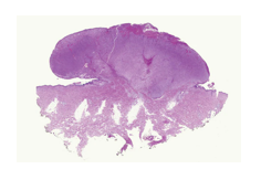
- Histopathological Features: Understanding cellular distribution, patterns of melanocyte nests, and the progression from benign naevi to malignant melanomas
- Prognostic Indicators: Measuring Breslow thickness, identifying Clark levels, and evaluating factors such as ulceration, mitotic activity, and lymphovascular invasion
- Diagnostic Challenges: Addressing issues in assessing margins, asymmetry, and single-cell spread in melanomas with complex presentations
- Case Studies: Real-world examples to illustrate atypical presentations, histological pitfalls, and misdiagnoses, emphasizing the importance of thorough evaluation and correlation with clinical findings
- Dermoscopy Integration: Applying low-power pattern recognition to enhance diagnostic accuracy, supported by examples of common and rare lesion subtypes, including spindle cell and balloon cell melanomas.
This course offers practical insights into managing uncertainties in histopathology, interpreting challenging cases, and collaborating effectively with dermatopathologists. By the end, participants will be adept at diagnosing melanocytic lesions, understanding their clinical implications, and guiding patient management with confidence.
Learning Outcomes
- Apply clinical and dermatoscopic information in evidence-based decision making with respect to skin lesions
- Define and use key terminology for dermatoscopy
- Apply decision algorithms in the diagnosis of both pigmented and non-pigmented skin lesions
- Define and use terminology used in dermatopathology
- Interpret the histological features of melanocytic lesions: lentigo simplex, naevi and melanomas.
Details
Cost: Please refer to Healthcert's website
Suitable for: All degree qualified medical practitioners
Study mode: 100% online
Disclaimer: Please note, once you click 'Register now' you will be leaving the AMA’s CPD Home website and entering a third-party education provider’s website. If you choose to register for this learning, you will need to provide some of your personal information directly to the third-party education provider. If you have any queries about how third-party education providers use, disclose or store your personal information you should consult their privacy policy.
Upon completion, your CPD activity record may take up to 4 weeks to be reflected on your CPD Home Dashboard.